
Artículos relacionados a Scanning Electron Microscopy and X-Ray Microanalysis:...
Sinopsis
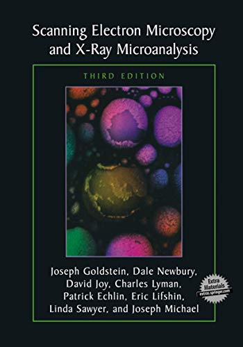
This text provides students as well as practitioners with a comprehensive introduction to the field of scanning electron microscopy (SEM) and X-ray microanalysis. The authors emphasize the practical aspects of the techniques described. Topics discussed include user-controlled functions of scanning electron microscopes and x-ray spectrometers and the use of x-rays for qualitative and quantitative analysis. Separate chapters cover SEM sample preparation methods for hard materials, polymers, and biological specimens. In addition techniques for the elimination of charging in non-conducting specimens are detailed.
"Sinopsis" puede pertenecer a otra edición de este libro.
De la contraportada
In the decade since the publication of the second edition of Scanning Electron Microscopy and X-Ray Microanalysis, there has been a great expansion in the capabilities of the basic scanning electron microscope (SEM) and the x-ray spectrometers. The emergence of the variab- pressure/environmental SEM has enabled the observation of samples c- taining water or other liquids or vapor and has allowed for an entirely new class of dynamic experiments, that of direct observation of che- cal reactions in situ. Critical advances in electron detector technology and computer-aided analysis have enabled structural (crystallographic) analysis of specimens at the micrometer scale through electron backscatter diffr- tion (EBSD). Low-voltage operation below 5 kV has improved x-ray spatial resolution by more than an order of magnitude and provided an effective route to minimizing sample charging. High-resolution imaging has cont- ued to develop with a more thorough understanding of how secondary el- trons are generated. The ?eld emission gun SEM, with its high brightness, advanced electron optics, which minimizes lens aberrations to yield an - fective nanometer-scale beam, and "through-the-lens" detector to enhance the measurement of primary-beam-excited secondary electrons, has made high-resolution imaging the rule rather than the exception. Methods of x-ray analysis have evolved allowing for better measurement of specimens with complex morphology: multiple thin layers of different compositions, and rough specimens and particles. Digital mapping has transformed classic x-ray area scanning, a purely qualitative technique, into fully quantitative compositional mapping.
"Sobre este título" puede pertenecer a otra edición de este libro.
Resultados de la búsqueda para Scanning Electron Microscopy and X-Ray Microanalysis:...

Imagen de archivo
Scanning Electron Microscopy and X-Ray Microanalysis: Third Edition
Librería: Emerald Green Media, Simi Valley, CA, Estados Unidos de America
Calificación del vendedor: 5 de 5 estrellas

paperback. Condición: Very Good. Clean Copy, May have light wear on cover/edges, otherwise very good! We've been in business for over 25 years serving thousands of customers worldwide. Your satisfaction is extremely important to us.Established Seller, We Ship Daily! We pack your order with care so that it arrives in great condition. Nº de ref. del artículo: mon0000060573
Comprar usado
EUR 50,77
Envío por EUR 4,02
Se envía dentro de Estados Unidos de America
Se envía dentro de Estados Unidos de America
Cantidad disponible: 1 disponibles
Imagen de archivo
Scanning Electron Microscopy and X-Ray Microanalysis: Third Edition
Librería: Marlton Books, Bridgeton, NJ, Estados Unidos de America
Calificación del vendedor: 5 de 5 estrellas
Condición: Acceptable. Readable, but has significant damage / tears. Has a remainder mark. paperback Used - Acceptable 2013. Nº de ref. del artículo: AB-022991
Comprar usado
EUR 53,53
Envío por EUR 2,69
Se envía dentro de Estados Unidos de America
Se envía dentro de Estados Unidos de America
Cantidad disponible: 1 disponibles
Imagen de archivo
Scanning Electron Microscopy and X-Ray Microanalysis (eng)
Impresión bajo demandaLibrería: Brook Bookstore On Demand, Napoli, NA, Italia
Calificación del vendedor: 5 de 5 estrellas
Condición: new. Questo è un articolo print on demand. Nº de ref. del artículo: V1FJBALJXL
Comprar nuevo
EUR 94,25
Envío por EUR 11,00
Se envía de Italia a Estados Unidos de America
Se envía de Italia a Estados Unidos de America
Cantidad disponible: Más de 20 disponibles
Imagen del vendedor
Scanning Electron Microscopy and X-Ray Microanalysis
Librería: GreatBookPrices, Columbia, MD, Estados Unidos de America
Calificación del vendedor: 5 de 5 estrellas
Condición: New. Nº de ref. del artículo: 21687683-n
Comprar nuevo
EUR 109,12
Envío por EUR 2,37
Se envía dentro de Estados Unidos de America
Se envía dentro de Estados Unidos de America
Cantidad disponible: Más de 20 disponibles
Imagen del vendedor
Scanning Electron Microscopy and X-Ray Microanalysis
Librería: GreatBookPricesUK, Woodford Green, Reino Unido
Calificación del vendedor: 5 de 5 estrellas
Condición: New. Nº de ref. del artículo: 21687683-n
Comprar nuevo
EUR 103,58
Envío por EUR 18,00
Se envía de Reino Unido a Estados Unidos de America
Se envía de Reino Unido a Estados Unidos de America
Cantidad disponible: Más de 20 disponibles
Imagen del vendedor
Scanning Electron Microscopy and X-Ray Microanalysis
Publicado por
Springer-Verlag New York Inc., US, 2013
ISBN 10: 1461349699
ISBN 13: 9781461349693
Nuevo
Paperback
Librería: Rarewaves.com USA, London, LONDO, Reino Unido
Calificación del vendedor: 5 de 5 estrellas
Paperback. Condición: New. Third Edition 2003. In the decade since the publication of the second edition of Scanning Electron Microscopy and X-Ray Microanalysis, there has been a great expansion in the capabilities of the basic scanning electron microscope (SEM) and the x-ray spectrometers. The emergence of the variab- pressure/environmental SEM has enabled the observation of samples c- taining water or other liquids or vapor and has allowed for an entirely new class of dynamic experiments, that of direct observation of che- cal reactions in situ. Critical advances in electron detector technology and computer-aided analysis have enabled structural (crystallographic) analysis of specimens at the micrometer scale through electron backscatter diffr- tion (EBSD). Low-voltage operation below 5 kV has improved x-ray spatial resolution by more than an order of magnitude and provided an effective route to minimizing sample charging. High-resolution imaging has cont- ued to develop with a more thorough understanding of how secondary el- trons are generated. The ?eld emission gun SEM, with its high brightness, advanced electron optics, which minimizes lens aberrations to yield an - fective nanometer-scale beam, and "through-the-lens" detector to enhance the measurement of primary-beam-excited secondary electrons, has made high-resolution imaging the rule rather than the exception. Methods of x-ray analysis have evolved allowing for better measurement of specimens with complex morphology: multiple thin layers of different compositions, and rough specimens and particles. Digital mapping has transformed classic x-ray area scanning, a purely qualitative technique, into fully quantitative compositional mapping. Nº de ref. del artículo: LU-9781461349693
Comprar nuevo
EUR 129,11
Gastos de envío gratis
Se envía de Reino Unido a Estados Unidos de America
Se envía de Reino Unido a Estados Unidos de America
Cantidad disponible: Más de 20 disponibles
Imagen de archivo
Scanning Electron Microscopy and X-Ray Microanalysis: Third Edition
Publicado por
Springer-Verlag New York Inc., 2013
ISBN 10: 1461349699
ISBN 13: 9781461349693
Nuevo
Paperback / softback
Librería: THE SAINT BOOKSTORE, Southport, Reino Unido
Calificación del vendedor: 5 de 5 estrellas
Paperback / softback. Condición: New. This item is printed on demand. New copy - Usually dispatched within 5-9 working days. Nº de ref. del artículo: C9781461349693
Comprar nuevo
EUR 110,32
Envío por EUR 27,97
Se envía de Reino Unido a Estados Unidos de America
Se envía de Reino Unido a Estados Unidos de America
Cantidad disponible: Más de 20 disponibles
Imagen del vendedor
Scanning Electron Microscopy and X-Ray Microanalysis
Publicado por
Springer US Mai 2013, 2013
ISBN 10: 1461349699
ISBN 13: 9781461349693
Nuevo
Taschenbuch
Librería: BuchWeltWeit Ludwig Meier e.K., Bergisch Gladbach, Alemania
Calificación del vendedor: 5 de 5 estrellas
Taschenbuch. Condición: Neu. This item is printed on demand - it takes 3-4 days longer - Neuware -This text provides students as well as practitioners with a comprehensive introduction to the field of scanning electron microscopy (SEM) and X-ray microanalysis. The authors emphasize the practical aspects of the techniques described. Topics discussed include user-controlled functions of scanning electron microscopes and x-ray spectrometers and the use of x-rays for qualitative and quantitative analysis. Separate chapters cover SEM sample preparation methods for hard materials, polymers, and biological specimens. In addition techniques for the elimination of charging in non-conducting specimens are detailed. 720 pp. Englisch. Nº de ref. del artículo: 9781461349693
Comprar nuevo
EUR 117,69
Envío por EUR 23,00
Se envía de Alemania a Estados Unidos de America
Se envía de Alemania a Estados Unidos de America
Cantidad disponible: 2 disponibles
Imagen de archivo
Scanning Electron Microscopy and X-Ray Microanalysis: Third Edition
Librería: California Books, Miami, FL, Estados Unidos de America
Calificación del vendedor: 5 de 5 estrellas
Condición: New. Nº de ref. del artículo: I-9781461349693
Comprar nuevo
EUR 141,23
Envío por EUR 2,69
Se envía dentro de Estados Unidos de America
Se envía dentro de Estados Unidos de America
Cantidad disponible: Más de 20 disponibles
Imagen del vendedor
Scanning Electron Microscopy and X-ray Microanalysis
Impresión bajo demandaLibrería: moluna, Greven, Alemania
Calificación del vendedor: 5 de 5 estrellas
Condición: New. Dieser Artikel ist ein Print on Demand Artikel und wird nach Ihrer Bestellung fuer Sie gedruckt. The text has been used in educating over 3,000 students at the Lehigh SEM short course as well as thousands of undergraduate and graduate students at universities in every corner of the globeThe authors have made extensive changes to the text and . Nº de ref. del artículo: 4193211
Comprar nuevo
EUR 101,04
Envío por EUR 48,99
Se envía de Alemania a Estados Unidos de America
Se envía de Alemania a Estados Unidos de America
Cantidad disponible: Más de 20 disponibles