
Importe total (1 artículo artículos):
Destino del pedido:

demian e s (20 resultados)
Ir a los resultados principales
Filtros de búsqueda
Tipo de artículo
- Todos los tipos de productos
- Libros (20)
- Revistas y publicaciones (No hay ningún otro resultado que coincida con este filtro.)
- Cómics (No hay ningún otro resultado que coincida con este filtro.)
- Partituras (No hay ningún otro resultado que coincida con este filtro.)
- Arte, grabados y pósters (No hay ningún otro resultado que coincida con este filtro.)
- Fotografías (No hay ningún otro resultado que coincida con este filtro.)
- Mapas (No hay ningún otro resultado que coincida con este filtro.)
- Manuscritos y coleccionismo de papel (No hay ningún otro resultado que coincida con este filtro.)
Condición Más información
- Nuevo (2)
- Como nuevo, Excelente o Muy bueno (No hay ningún otro resultado que coincida con este filtro.)
- Bueno o Aceptable (No hay ningún otro resultado que coincida con este filtro.)
- Regular o Pobre (No hay ningún otro resultado que coincida con este filtro.)
- Tal como se indica (18)
Encuadernación
- Todas
- Tapa dura (No hay ningún otro resultado que coincida con este filtro.)
- Tapa blanda (2)
Más atributos
- Primera edición (No hay ningún otro resultado que coincida con este filtro.)
- Firmado (No hay ningún otro resultado que coincida con este filtro.)
- Sobrecubierta (No hay ningún otro resultado que coincida con este filtro.)
- Con imágenes (14)
- No impresión bajo demanda (20)
Idioma (2)
Precio
- Cualquier precio
- Menos de EUR 20
- EUR 20 a EUR 45
- Más de EUR 45 (No hay ningún otro resultado que coincida con este filtro.)
Gastos de envío gratis
- Envío gratis a Estados Unidos de America (No hay ningún otro resultado que coincida con este filtro.)
Ubicación del vendedor
Valoración de los vendedores
-

El rostro oculto del animal
Librería: KALAMO BOOKS, Burriana, CS, Espańa
Calificación del vendedor: 5 de 5 estrellas

EUR 11,50
Envío por EUR 17,86
Se envía de Espańa a Estados Unidos de AmericaCantidad disponible: 1 disponibles
Ańadir al carritoTapa blanda. Condición: Nuevo.
-

ANTOLOGĂA POĂ?TICA. ManĂas del corazĂłn
Idioma: Espańol
Publicado por GRUPO EDITORIAL LETRAS NEGRAS S.A.S
ISBN 10: 6287680156 ISBN 13: 9786287680159
Librería: KALAMO BOOKS, Burriana, CS, Espańa
Calificación del vendedor: 5 de 5 estrellas
EUR 13,50
Envío por EUR 17,86
Se envía de Espańa a Estados Unidos de AmericaCantidad disponible: 1 disponibles
Ańadir al carritoTapa blanda. Condición: Nuevo.
-

The egg mass, egg laying and mating behaviour of the snail Marisa cornuarietis (L.)
Publicado por 1970-1971, 1970
Librería: ConchBooks, Harxheim, Alemania
Calificación del vendedor: 4 de 5 estrellas

EUR 5,75
Envío por EUR 24,00
Se envía de Alemania a Estados Unidos de AmericaCantidad disponible: 1 disponibles
Ańadir al carrito12 pp., 3 figs, gr. 8.
-
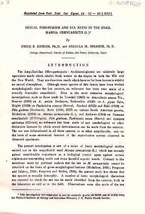
Sexual dimorphism and sex ratio in the snail Marisa cornuarietis (L.)
Ańo de publicación: 1972
Librería: ConchBooks, Harxheim, Alemania
Calificación del vendedor: 4 de 5 estrellas
EUR 5,75
Envío por EUR 24,00
Se envía de Alemania a Estados Unidos de AmericaCantidad disponible: 1 disponibles
Ańadir al carrito12 pp., 5 figs, gr. 8.
-

Prospects of the use of Marisa cornuarietis in the biological control of Limnaea cailliaudi in th U.A.R
Ańo de publicación: 1965
Librería: ConchBooks, Harxheim, Alemania
Calificación del vendedor: 4 de 5 estrellas
EUR 5,75
Envío por EUR 24,00
Se envía de Alemania a Estados Unidos de AmericaCantidad disponible: 1 disponibles
Ańadir al carrito5 pp., 3 pls, gr. 8.
-

Predatory activity of Marisa cornuarietis against Bulinus (Bulinus) truncatus, the transmitter of urinary schistosomiasis
Ańo de publicación: 1965
Librería: ConchBooks, Harxheim, Alemania
Calificación del vendedor: 4 de 5 estrellas
EUR 5,75
Envío por EUR 24,00
Se envía de Alemania a Estados Unidos de AmericaCantidad disponible: 1 disponibles
Ańadir al carrito6 pp. + 4 pls, gr. 8.
-
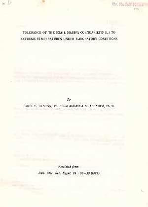
Tolerance of the snail Marisa cornuarietis (L.) to extreme temperature under laboratory conditions
Ańo de publicación: 1972
Librería: ConchBooks, Harxheim, Alemania
Calificación del vendedor: 4 de 5 estrellas
EUR 5,75
Envío por EUR 24,00
Se envía de Alemania a Estados Unidos de AmericaCantidad disponible: 1 disponibles
Ańadir al carrito11 pp., 4 figs, gr. 8.
-

Embryonic development and organogenesis in the snail Marisa cornuarietis (Mesogastropoda: Ampullariidae). V. Development of the nervous system
Ańo de publicación: 1975
Librería: ConchBooks, Harxheim, Alemania
Calificación del vendedor: 4 de 5 estrellas
EUR 5,75
Envío por EUR 24,00
Se envía de Alemania a Estados Unidos de AmericaCantidad disponible: 1 disponibles
Ańadir al carritoThe nervous system is ectodermal in origin. All nerve ganglia arise separately by proliferation and later delamination from the ectoderm, not by invagination. They become secondarily connected to one another by commissures and connectives developing as extensions from the peripheral layer of ganglionic nerve cells. Rudiments of the cerebral, pedal, pleural and intestinal (parietal) ganglia arise almost simultaneously at a relatively early stage (Stage V). The cerebral ganglia develop from the ectoderm of the head plates. Rudiments of the pedal and pleural ganglia are separate at their inception. They later fuse (Stage VI) to form a pleuro-pedal ganglionic mass on each side. The 2 intestinal ganglia are symmetrical at the beginning, but they soon lose their symmetry as a result of torsion. The right ganglion crosses to the left over the gut and persists as the supraintestinal ganglion. The left or subintestinal ganglion shifts to the right and forward, and fuses with the right pleural ganglion (Stage VIII), thus obscuring the chiastoneury. The paired buccal and single visceral (abdominal) ganglia start differentiating in Stage VII. The former develop from the ectodermal wall of the stomodaeum, while the visceral ganglion delaminates from the right wall of the visceral sac, then shifts to the left during torsion. The statocysts develop early (Stage V) from 2 ectodermal invaginations on either side of the rudimentary foot. They later separate from the overlying ectoderm and statoconi appear in their lumina. Contrary to earlier reports on related ampullariids, the osphradium proved to be ontogenetically older than the mantle and mantle cavity. It starts differentiating as a thickened ectodermal plate in the right wall of the visceral sac (Stage V). During torsion, it becomes engulfed in the mantle cavity and shifts to the left side, then is carried forward as the mantlegrow. The eyes develop late (Stage IX) as ectodermal invaginations which rapidly separate from the ectoderm to form closed vesicles. Their cells start differentiating before hatching to form the retina, in which pigment is deposited, and the inner cornea. The lens is secreted in the lumen of the eye and grows by addition of concentric layers of secretion. 14 pp., 10 figs, gr. 8.
-

Studies on the Chromosome Numbers in the Ampulariidae (Gastropoda, Prosobranchia)
Ańo de publicación: 1965
Librería: ConchBooks, Harxheim, Alemania
Calificación del vendedor: 4 de 5 estrellas
EUR 5,75
Envío por EUR 24,00
Se envía de Alemania a Estados Unidos de AmericaCantidad disponible: 1 disponibles
Ańadir al carritoTreated taxa: Marisa cornuarietis, Lanistes bolteni, Pila ovata 6 pp. + 3 pls, 4.
-

Embryonic development and organogenesis in the snail Marisa cornuarietis (Mesogastropoda: Ampullariidae). IV. Development of the shell gland, mantle and respiratory organs
Ańo de publicación: 1973
Librería: ConchBooks, Harxheim, Alemania
Calificación del vendedor: 4 de 5 estrellas
EUR 5,87
Envío por EUR 24,00
Se envía de Alemania a Estados Unidos de AmericaCantidad disponible: 1 disponibles
Ańadir al carrito17 pp., 13 figs, gr. 8.
-

Embryonic development and organogenesis in the snail Marisa cornuarietis (Mesogastropoda: Ampullariidae). III. Development of the circulatory and renal system
Ańo de publicación: 1973
Librería: ConchBooks, Harxheim, Alemania
Calificación del vendedor: 4 de 5 estrellas
EUR 6,90
Envío por EUR 24,00
Se envía de Alemania a Estados Unidos de AmericaCantidad disponible: 1 disponibles
Ańadir al carritoThe nervous system is ectodermal in origin. All nerve ganglia arise separately by proliferation and later delamination from the ectoderm, not by invagination. They become secondarily connected to one another by commissures and connectives developing as extensions from the peripheral layer of ganglionic nerve cells. Rudiments of the cerebral, pedal, pleural and intestinal (parietal) ganglia arise almost simultaneously at a relatively early stage (Stage V). The cerebral ganglia develop from the ectoderm of the head plates. Rudiments of the pedal and pleural ganglia are separate at their inception. They later fuse (Stage VI) to form a pleuro-pedal ganglionic mass on each side. The 2 intestinal ganglia are symmetrical at the beginning, but they soon lose their symmetry as a result of torsion. The right ganglion crosses to the left over the gut and persists as the supraintestinal ganglion. The left or subintestinal ganglion shifts to the right and forward, and fuses with the right pleural ganglion (Stage VIII), thus obscuring the chiastoneury. The paired buccal and single visceral (abdominal) ganglia start differentiating in Stage VII. The former develop from the ectodermal wall of the stomodaeum, while the visceral ganglion delaminates from the right wall of the visceral sac, then shifts to the left during torsion. The statocysts develop early (Stage V) from 2 ectodermal invaginations on either side of the rudimentary foot. They later separate from the overlying ectoderm and statoconi appear in their lumina. Contrary to earlier reports on related ampullariids, the osphradium proved to be ontogenetically older than the mantle and mantle cavity. It starts differentiating as a thickened ectodermal plate in the right wall of the visceral sac (Stage V). During torsion, it becomes engulfed in the mantle cavity and shifts to the left side, then is carried forward as the mantlegrow. The eyes develop late (Stage IX) as ectodermal invaginations which rapidly separate from the ectoderm to form closed vesicles. Their cells start differentiating before hatching to form the retina, in which pigment is deposited, and the inner cornea. The lens is secreted in the lumen of the eye and grows by addition of concentric layers of secretion. 20 pp., 10 figs, gr. 8.
-
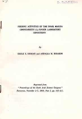
Feeding activities of the snail Marisa cornuarietis (L.) under laboratory conditions
Ańo de publicación: 1969
Librería: ConchBooks, Harxheim, Alemania
Calificación del vendedor: 4 de 5 estrellas
EUR 7,25
Envío por EUR 24,00
Se envía de Alemania a Estados Unidos de AmericaCantidad disponible: 1 disponibles
Ańadir al carrito21 pp., 2 figs, gr. 8.
-

The histology of the respiratory organs of Marisa cornuarietis (L.)
Ańo de publicación: 1965
Librería: ConchBooks, Harxheim, Alemania
Calificación del vendedor: 4 de 5 estrellas
EUR 7,25
Envío por EUR 24,00
Se envía de Alemania a Estados Unidos de AmericaCantidad disponible: 1 disponibles
Ańadir al carrito21 pp., 9 figs, stapled gr. 8.
-

Embryonic development and organogenesis in the snail Marisa cornuarietis (Mesogastropoda: Ampullariidae). II. Development of the alimentary system
Ańo de publicación: 1973
Librería: ConchBooks, Harxheim, Alemania
Calificación del vendedor: 4 de 5 estrellas
EUR 8,28
Envío por EUR 24,00
Se envía de Alemania a Estados Unidos de AmericaCantidad disponible: 1 disponibles
Ańadir al carrito24 pp., 16 figs, gr. 8.
-
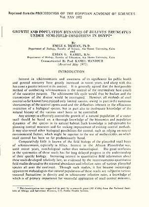
Growth and population dynamics of Bulinus truncatus under semi-field conditions in Egypt
Ańo de publicación: 1972
Librería: ConchBooks, Harxheim, Alemania
Calificación del vendedor: 4 de 5 estrellas
EUR 8,28
Envío por EUR 24,00
Se envía de Alemania a Estados Unidos de AmericaCantidad disponible: 1 disponibles
Ańadir al carrito24 pp., 13 figs, gr. 8.
-
Prospects of the use of Marisa cornuarietis in the biological control of Limnaea caillaudi in the U.A.R. In 4°, offp., pp. 8 with 3 pls. Offprint from Proc. Egyptian Ac. Sci., 18
Ańo de publicación: 1965
Librería: Riccardo Giannuzzi Savelli, Palermo, PA, Italia
Calificación del vendedor: 2 de 5 estrellas

EUR 3,50
Envío por EUR 30,00
Se envía de Italia a Estados Unidos de AmericaCantidad disponible: 1 disponibles
Ańadir al carrito -

Embryonic development and organogenesis in the snail Marisa cornuarietis (Mesogastropoda: Ampullariidae). I. General outlines of development
Ańo de publicación: 1973
Librería: ConchBooks, Harxheim, Alemania
Calificación del vendedor: 4 de 5 estrellas
EUR 9,66
Envío por EUR 24,00
Se envía de Alemania a Estados Unidos de AmericaCantidad disponible: 1 disponibles
Ańadir al carrito28 pp., 24 figs, gr. 8.
-

The histology of the alimentary system of Marisa cornuarietis (Mesogastropoda: Ampullariidae)
Ańo de publicación: 1967
Librería: ConchBooks, Harxheim, Alemania
Calificación del vendedor: 4 de 5 estrellas
EUR 9,89
Envío por EUR 24,00
Se envía de Alemania a Estados Unidos de AmericaCantidad disponible: 1 disponibles
Ańadir al carrito48 pp., 59 figs, gr. 8.
-
Embryonic development and organogenesis in the snail Marisa cornuarietis (Mesogastropoda: Ampullariidae) - II Development of the alimentary system. In 8vo, offp., pp. 24 with 16 figs. Offprint from Malacologia 12(1)
Ańo de publicación: 1973
Librería: Riccardo Giannuzzi Savelli, Palermo, PA, Italia
Calificación del vendedor: 2 de 5 estrellas
EUR 4,00
Envío por EUR 30,00
Se envía de Italia a Estados Unidos de AmericaCantidad disponible: 1 disponibles
Ańadir al carrito -
Collection of 17 papers on anatomy, physiology and development of Marisa cornuarietis.
Publicado por 1964-1975, 1964
Librería: Hermann L. Strack, Loguivy Plougras, Francia
Calificación del vendedor: 5 de 5 estrellas
EUR 38,50
Envío por EUR 48,00
Se envía de Francia a Estados Unidos de AmericaCantidad disponible: 1 disponibles
Ańadir al carritoAll are stapled or in printed wrappers. Some quite large like ??Anatomy of the alimentary system of marisa cornuarietis?? (75 p., 21 figs). All ex library Dr. A.C. van Bruggen (with his stamp).
