
Importe total (1 artículo artículos):
Destino del pedido:

9781626239845 - Imaging Anatomy: Text and Atlas Volume 3: Bones, Joints, Muscles, Vessels, and Nerves (Atlas of Imaging Anatomy) de Saremi, Farhood; Patel, Dakshesh; Sanchez-Quinta... (18 resultados)
Ir a los resultados principales
Filtros de búsqueda
Tipo de artículo
- Todos los tipos de productos
- Libros (18)
- Revistas y publicaciones (No hay ningún otro resultado que coincida con este filtro.)
- Cómics (No hay ningún otro resultado que coincida con este filtro.)
- Partituras (No hay ningún otro resultado que coincida con este filtro.)
- Arte, grabados y pósters (No hay ningún otro resultado que coincida con este filtro.)
- Fotografías (No hay ningún otro resultado que coincida con este filtro.)
- Mapas (No hay ningún otro resultado que coincida con este filtro.)
- Manuscritos y coleccionismo de papel (No hay ningún otro resultado que coincida con este filtro.)
Condición Más información
- Nuevo (16)
- Como nuevo, Excelente o Muy bueno (2)
- Bueno o Aceptable (No hay ningún otro resultado que coincida con este filtro.)
- Regular o Pobre (No hay ningún otro resultado que coincida con este filtro.)
- Tal como se indica (No hay ningún otro resultado que coincida con este filtro.)
Encuadernación
- Todas
- Tapa dura (18)
- Tapa blanda (No hay ningún otro resultado que coincida con este filtro.)
Más atributos
- Primera edición (1)
- Firmado (No hay ningún otro resultado que coincida con este filtro.)
- Sobrecubierta (No hay ningún otro resultado que coincida con este filtro.)
- Con imágenes (2)
- No impresión bajo demanda (18)
Idioma (1)
Precio
- Cualquier precio
- Menos de EUR 20 (No hay ningún otro resultado que coincida con este filtro.)
- EUR 20 a EUR 45 (No hay ningún otro resultado que coincida con este filtro.)
- Más de EUR 45
Gastos de envío gratis
Ubicación del vendedor
Valoración de los vendedores
-

Imaging Anatomy - Text and Atlas : Bones, Joints, Muscles, Vessels, and Nerves
Libro 3 de 3: Atlas of Imaging AnatomyLibrería: GreatBookPricesUK, Woodford Green, Reino Unido
Calificación del vendedor: 5 de 5 estrellas

EUR 165,68
Envío por EUR 17,28
Se envía de Reino Unido a Estados Unidos de AmericaCantidad disponible: 2 disponibles
Ańadir al carritoCondición: New.
-
Imaging Anatomy: Text and Atlas Volume 3
Libro 3 de 3: Atlas of Imaging AnatomyLibrería: PBShop.store UK, Fairford, GLOS, Reino Unido
Calificación del vendedor: 5 de 5 estrellas
EUR 165,69
Envío por EUR 22,78
Se envía de Reino Unido a Estados Unidos de AmericaCantidad disponible: 1 disponibles
Ańadir al carritoUNK. Condición: New. New Book. Shipped from UK. Established seller since 2000.
-
Imaging Anatomy - Text and Atlas : Bones, Joints, Muscles, Vessels, and Nerves
Libro 3 de 3: Atlas of Imaging AnatomyLibrería: GreatBookPrices, Columbia, MD, Estados Unidos de America
Calificación del vendedor: 5 de 5 estrellas
EUR 191,75
Envío por EUR 2,30
Se envía dentro de Estados Unidos de AmericaCantidad disponible: 2 disponibles
Ańadir al carritoCondición: New.
-
Imaging Anatomy - Text and Atlas : Bones, Joints, Muscles, Vessels, and Nerves
Libro 3 de 3: Atlas of Imaging AnatomyLibrería: GreatBookPrices, Columbia, MD, Estados Unidos de America
Calificación del vendedor: 5 de 5 estrellas
EUR 191,98
Envío por EUR 2,30
Se envía dentro de Estados Unidos de AmericaCantidad disponible: 2 disponibles
Ańadir al carritoCondición: As New. Unread book in perfect condition.
-
Imaging Anatomy - Text and Atlas : Bones, Joints, Muscles, Vessels, and Nerves
Libro 3 de 3: Atlas of Imaging AnatomyLibrería: GreatBookPricesUK, Woodford Green, Reino Unido
Calificación del vendedor: 5 de 5 estrellas
EUR 181,80
Envío por EUR 17,28
Se envía de Reino Unido a Estados Unidos de AmericaCantidad disponible: 2 disponibles
Ańadir al carritoCondición: As New. Unread book in perfect condition.
-
Imaging Anatomy: Text and Atlas Volume 3: Bones, Joints, Muscles, Vessels, and Nerves (Atlas of Imaging Anatomy)
Libro 3 de 3: Atlas of Imaging AnatomyLibrería: Russell Books, Victoria, BC, Canada
Calificación del vendedor: 5 de 5 estrellas
EUR 183,32
Envío por EUR 17,44
Se envía de Canada a Estados Unidos de AmericaCantidad disponible: 1 disponibles
Ańadir al carritohardcover. Condición: New. Special order direct from the distributor.
-
Atlas of Imaging Anatomy: Bones, Muscles, and Extremities
Libro 3 de 3: Atlas of Imaging AnatomyIdioma: Inglés
Publicado por Thieme Publishers New York, 2024
ISBN 10: 1626239843 ISBN 13: 9781626239845
Librería: moluna, Greven, Alemania
Calificación del vendedor: 5 de 5 estrellas
EUR 157,55
Envío por EUR 48,99
Se envía de Alemania a Estados Unidos de AmericaCantidad disponible: 5 disponibles
Ańadir al carritoCondición: New.
-
Imaging Anatomy: Text and Atlas Volume 3 (Hardcover)
Libro 3 de 3: Atlas of Imaging AnatomyIdioma: Inglés
Publicado por Thieme Medical Publishers Inc, New York, 2024
ISBN 10: 1626239843 ISBN 13: 9781626239845
Librería: Grand Eagle Retail, Bensenville, IL, Estados Unidos de America
Calificación del vendedor: 5 de 5 estrellas
EUR 220,30
Gastos de envío gratis
Se envía dentro de Estados Unidos de AmericaCantidad disponible: 1 disponibles
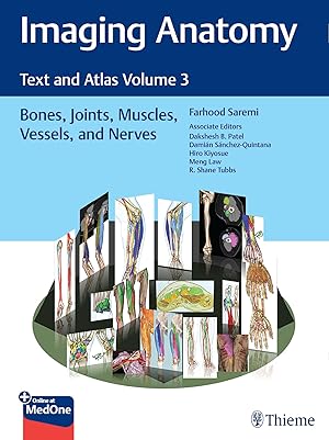
Ańadir al carritoHardcover. Condición: new. Hardcover. An in-depth guide to upper and lower extremity anatomy based on the latest imaging techniquesWhile the study of anatomy plays a fundamental role in the practice of medicine, most textbooks don't rely on modern imaging and post-processing methods to depict and increase its understanding. Imaging Anatomy Text and Atlas Volume 3: Bones, Joints, Muscles, Vessels, and Nerves is the third in a series of four richly illustrated radiologic references edited by distinguished radiologist Farhood Saremi. The atlas is coedited by esteemed colleagues Dakshesh B. Patel, Damian Sanchez-Quintana, Hiro Kiyosue, Meng Law, and R. Shane Tubbs and features contributions from an impressive group of international experts. The succinctly written text and superb images fill a gap in the literature, with descriptions of relevant anatomical components in the context of current advances in imaging technology and science.This exquisitely crafted atlas combines fundamental core anatomy principles with modern imaging and post-processing methods to increase understanding of intricate anatomical features. Twenty-four concise chapters cover terminology and classification of musculoskeletal structure, bones, muscles, joints, arteries, veins, nerves, and lymphatics. High-quality dissecting imaging anatomy, discussion of anatomical variants, postsurgical anatomy, and important pathology examples provide a strong foundation for differentiating normal versus pathologic anatomy.Key HighlightsState-of-the-art CT, MR, angiography, and ultrasound techniques infused with 3D reformations, color coded volume rendering, and 3-7 Tesla MR views delineate anatomy in great detailCross-sectional and topographic cadaveric views and illustrations by world-renowned anatomists improve the ability to grasp difficult radiology conceptsConsistently formatted chapters including an introduction, embryology, review of anatomy, discussion of anatomical variants, surgical anatomy, and congenital and acquired pathologies enhance learningThis unique atlas provides a virtual, user-friendly dissection experience, making it a must-have reference for medical students, radiology residents and veteran radiologists, internists, and general surgeons, as well as vascular and transplant surgeons. This book includes complimentary access to a digital copy on Publisher's Note: Products purchased from Third Party sellers are not guaranteed by the publisher for quality, authenticity, or access to any online entitlements included with the product. Blending core anatomical principles with modern imaging techniques, this richly illustrated atlas details CT, MR, angiography, and ultrasound views of bones, muscles, joints, vessels, and nerves. Cross?sectional and cadaveric images along with discussions of variants and pathologies aid in distinguishing normal from abnormal anatomy. Shipping may be from multiple locations in the US or from the UK, depending on stock availability.
-
Imaging Anatomy: Text and Atlas Volume 3: Bones, Joints, Muscles, Vessels, and Nerves (Atlas of Imaging Anatomy)
Libro 3 de 3: Atlas of Imaging AnatomyLibrería: Books Puddle, New York, NY, Estados Unidos de America
Calificación del vendedor: 4 de 5 estrellas

EUR 218,71
Envío por EUR 3,48
Se envía dentro de Estados Unidos de AmericaCantidad disponible: 4 disponibles
Ańadir al carritoCondición: New. 1st edition NO-PA16APR2015-KAP.
-
Imaging Anatomy: Text and Atlas Volume 3: Bones, Joints, Muscles, Vessels, and Nerves (Atlas of Imaging Anatomy)
Libro 3 de 3: Atlas of Imaging AnatomyLibrería: Majestic Books, Hounslow, Reino Unido
Calificación del vendedor: 4 de 5 estrellas
EUR 215,91
Envío por EUR 7,49
Se envía de Reino Unido a Estados Unidos de AmericaCantidad disponible: 4 disponibles
Ańadir al carritoCondición: New.
-
Atlas Of Imaging Anatomy Bones Muscle
Libro 3 de 3: Atlas of Imaging AnatomyIdioma: Inglés
Publicado por Thieme Medical Publishers, 2024
ISBN 10: 1626239843 ISBN 13: 9781626239845
Librería: Kennys Bookshop and Art Galleries Ltd., Galway, GY, Irlanda
Calificación del vendedor: 5 de 5 estrellas
Original o primera edición
EUR 215,66
Envío por EUR 10,50
Se envía de Irlanda a Estados Unidos de AmericaCantidad disponible: 1 disponibles
Ańadir al carritoCondición: New. 2024. 1st Edition. hardcover. . . . . .
-
Imaging Anatomy: Text and Atlas Volume 3: Bones, Joints, Muscles, Vessels, and Nerves (Atlas of Imaging Anatomy)
Libro 3 de 3: Atlas of Imaging AnatomyLibrería: Biblios, Frankfurt am main, HESSE, Alemania
Calificación del vendedor: 4 de 5 estrellas
EUR 217,42
Envío por EUR 9,95
Se envía de Alemania a Estados Unidos de AmericaCantidad disponible: 4 disponibles
Ańadir al carritoCondición: New.
-

Imaging Anatomy: Text and Atlas Volume 3 : Bones, Joints, Muscles, Vessels, and Nerves
Libro 3 de 3: Atlas of Imaging AnatomyIdioma: Inglés
Publicado por Thieme, Stuttgart Mär 2023, 2023
ISBN 10: 1626239843 ISBN 13: 9781626239845
Librería: AHA-BUCH GmbH, Einbeck, Alemania
Calificación del vendedor: 5 de 5 estrellas
EUR 187,22
Envío por EUR 83,59
Se envía de Alemania a Estados Unidos de AmericaCantidad disponible: 1 disponibles
Ańadir al carritoKombiprodukt. Condición: Neu. Neuware.
-
Imaging Anatomy: Text and Atlas Volume 3 Bones, Joints, Muscles, Vessels, and Nerves
Libro 3 de 3: Atlas of Imaging AnatomyLibrería: Revaluation Books, Exeter, Reino Unido
Calificación del vendedor: 5 de 5 estrellas
EUR 245,63
Envío por EUR 34,57
Se envía de Reino Unido a Estados Unidos de AmericaCantidad disponible: 1 disponibles
Ańadir al carritoHardcover. Condición: Brand New. 1st edition. 942 pages. 12.00x9.00x2.25 inches. In Stock.
-
Atlas Of Imaging Anatomy Bones Muscle
Libro 3 de 3: Atlas of Imaging AnatomyIdioma: Inglés
Publicado por Thieme Medical Publishers, 2024
ISBN 10: 1626239843 ISBN 13: 9781626239845
Librería: Kennys Bookstore, Olney, MD, Estados Unidos de America
Calificación del vendedor: 5 de 5 estrellas
EUR 279,98
Envío por EUR 9,16
Se envía dentro de Estados Unidos de AmericaCantidad disponible: 1 disponibles
Ańadir al carritoCondición: New. 2024. 1st Edition. hardcover. . . . . . Books ship from the US and Ireland.
-
Imaging Anatomy: Text and Atlas Volume 3 Bones, Joints, Muscles, Vessels, and Nerves
Libro 3 de 3: Atlas of Imaging AnatomyLibrería: Revaluation Books, Exeter, Reino Unido
Calificación del vendedor: 5 de 5 estrellas
EUR 310,36
Envío por EUR 34,57
Se envía de Reino Unido a Estados Unidos de AmericaCantidad disponible: 1 disponibles
Ańadir al carritoHardcover. Condición: Brand New. 1st edition. 942 pages. 12.00x9.00x2.25 inches. In Stock.
-
Imaging Anatomy: Text and Atlas Volume 3: Bones, Joints, Muscles, Vessels, and Nerves (Atlas of Imaging Anatomy)
Libro 3 de 3: Atlas of Imaging AnatomyLibrería: GoldBooks, Denver, CO, Estados Unidos de America
Calificación del vendedor: 5 de 5 estrellas
EUR 345,32
Envío por EUR 4,80
Se envía dentro de Estados Unidos de AmericaCantidad disponible: 1 disponibles
Ańadir al carritoHardcover. Condición: new. New Copy. Customer Service Guaranteed.
-
Imaging Anatomy: Text and Atlas Volume 3 (Hardcover)
Libro 3 de 3: Atlas of Imaging AnatomyIdioma: Inglés
Publicado por Thieme Medical Publishers Inc, New York, 2024
ISBN 10: 1626239843 ISBN 13: 9781626239845
Librería: AussieBookSeller, Truganina, VIC, Australia
Calificación del vendedor: 5 de 5 estrellas
EUR 444,64
Envío por EUR 32,28
Se envía de Australia a Estados Unidos de AmericaCantidad disponible: 1 disponibles
Ańadir al carritoHardcover. Condición: new. Hardcover. An in-depth guide to upper and lower extremity anatomy based on the latest imaging techniquesWhile the study of anatomy plays a fundamental role in the practice of medicine, most textbooks don't rely on modern imaging and post-processing methods to depict and increase its understanding. Imaging Anatomy Text and Atlas Volume 3: Bones, Joints, Muscles, Vessels, and Nerves is the third in a series of four richly illustrated radiologic references edited by distinguished radiologist Farhood Saremi. The atlas is coedited by esteemed colleagues Dakshesh B. Patel, Damian Sanchez-Quintana, Hiro Kiyosue, Meng Law, and R. Shane Tubbs and features contributions from an impressive group of international experts. The succinctly written text and superb images fill a gap in the literature, with descriptions of relevant anatomical components in the context of current advances in imaging technology and science.This exquisitely crafted atlas combines fundamental core anatomy principles with modern imaging and post-processing methods to increase understanding of intricate anatomical features. Twenty-four concise chapters cover terminology and classification of musculoskeletal structure, bones, muscles, joints, arteries, veins, nerves, and lymphatics. High-quality dissecting imaging anatomy, discussion of anatomical variants, postsurgical anatomy, and important pathology examples provide a strong foundation for differentiating normal versus pathologic anatomy.Key HighlightsState-of-the-art CT, MR, angiography, and ultrasound techniques infused with 3D reformations, color coded volume rendering, and 3-7 Tesla MR views delineate anatomy in great detailCross-sectional and topographic cadaveric views and illustrations by world-renowned anatomists improve the ability to grasp difficult radiology conceptsConsistently formatted chapters including an introduction, embryology, review of anatomy, discussion of anatomical variants, surgical anatomy, and congenital and acquired pathologies enhance learningThis unique atlas provides a virtual, user-friendly dissection experience, making it a must-have reference for medical students, radiology residents and veteran radiologists, internists, and general surgeons, as well as vascular and transplant surgeons. This book includes complimentary access to a digital copy on Publisher's Note: Products purchased from Third Party sellers are not guaranteed by the publisher for quality, authenticity, or access to any online entitlements included with the product. Blending core anatomical principles with modern imaging techniques, this richly illustrated atlas details CT, MR, angiography, and ultrasound views of bones, muscles, joints, vessels, and nerves. Cross?sectional and cadaveric images along with discussions of variants and pathologies aid in distinguishing normal from abnormal anatomy. Shipping may be from our Sydney, NSW warehouse or from our UK or US warehouse, depending on stock availability.