Artículos relacionados a Color Atlas of Anatomy - international edition: A Photograph...
Color Atlas of Anatomy - international edition: A Photographic Study of the Human Body - Tapa blanda

- EditorialSchattauer Gmbh (2015-01-29)
- Ańo de publicación1656
- ISBN 10 3794529820
- ISBN 13 9783794529827
- EncuadernaciónTapa blanda
Comprar nuevo
Ver este artículo
EUR 106,33
Gastos de envío:
EUR 2,47
A Estados Unidos de America
Los mejores resultados en AbeBooks
Imagen de archivo
Color Atlas of Anatomy - international edition
Publicado por
Schattauer GmbH
(2015)
ISBN 10: 3794529820
ISBN 13: 9783794529827
Nuevo
Tapa blanda
Cantidad disponible: 3
Librería:
Valoración
Descripción Condición: New. Nş de ref. del artículo: 23676082-n
Comprar nuevo
EUR 106,33
Convertir moneda
Imagen de archivo
Color Atlas of Anatomy international edition A Photographic Study of the Human Body
Publicado por
Schattauer GmbH
(2015)
ISBN 10: 3794529820
ISBN 13: 9783794529827
Nuevo
PAP
Cantidad disponible: 2
Librería:
Valoración
Descripción PAP. Condición: New. New Book. Shipped from UK. Established seller since 2000. Nş de ref. del artículo: DB-9783794529827
Comprar nuevo
EUR 108,88
Convertir moneda
Imagen de archivo
Color Atlas of Anatomy - international edition
Publicado por
Schattauer GmbH
(2015)
ISBN 10: 3794529820
ISBN 13: 9783794529827
Nuevo
Tapa blanda
Cantidad disponible: 3
Librería:
Valoración
Descripción Condición: New. Nş de ref. del artículo: 23676082-n
Comprar nuevo
EUR 94,19
Convertir moneda
Gastos de envío:
EUR 17,53
De Reino Unido a Estados Unidos de America
Destinos, gastos y plazos de envío
Imagen del vendedor
Color Atlas of Anatomy - international edition
Publicado por
Schattauer Gmbh Jan 2015
(2015)
ISBN 10: 3794529820
ISBN 13: 9783794529827
Nuevo
Taschenbuch
Cantidad disponible: 2
Librería:
Valoración
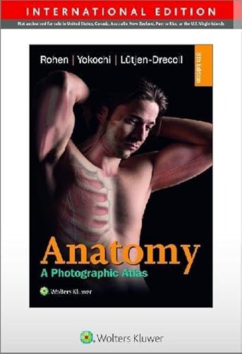
Descripción Taschenbuch. Condición: Neu. Neuware -Prepare for the dissection lab and operating room with this proven atlas.Featuring outstanding full-color photographs of actual cadaver dissections with accompanying schematic drawings and diagnostic images, Anatomy: A Photographic Atlas depicts anatomic structures more realistically than illustrations in traditional atlases. Chapters are organized by region in the order of a typical dissection with each chapter presenting topographical anatomical structures in a systemic manner.- Authentic photographic reproduction of colors, structures, and spatial dimensions as seen in the dissection lab and on the operating table help you develop an understanding of the anatomy of the human body.- Functional connections between single organs, the surrounding tissue, and organ systems are clarified to prepare you for the dissection lab and practical exams.- Clinical cases and over 1,200 images enhance your understanding.- Dissections illustrate the topographical anatomy in layers 'from the outside in' to better prepare you for the lab and operating room.New to the 8th edition:- Additional images including clinical imaging (MRIs, CTs, and endoscopic techniques).- A more modern and cohesive art program includes new modern MRI images as well as new full-color dissection photographs that replace black-and-white dissection images.- Introductory pages to each chapter have been redesigned for more clarity. 532 pp. Deutsch. Nş de ref. del artículo: 9783794529827
Comprar nuevo
EUR 96,99
Convertir moneda
Gastos de envío:
EUR 17,13
De Alemania a Estados Unidos de America
Destinos, gastos y plazos de envío
Imagen del vendedor
Color Atlas of Anatomy - international edition
Publicado por
Schattauer Gmbh Jan 2015
(2015)
ISBN 10: 3794529820
ISBN 13: 9783794529827
Nuevo
Taschenbuch
Cantidad disponible: 2
Librería:
Valoración
Descripción Taschenbuch. Condición: Neu. Neuware -Prepare for the dissection lab and operating room with this proven atlas.Featuring outstanding full-color photographs of actual cadaver dissections with accompanying schematic drawings and diagnostic images, Anatomy: A Photographic Atlas depicts anatomic structures more realistically than illustrations in traditional atlases. Chapters are organized by region in the order of a typical dissection with each chapter presenting topographical anatomical structures in a systemic manner.- Authentic photographic reproduction of colors, structures, and spatial dimensions as seen in the dissection lab and on the operating table help you develop an understanding of the anatomy of the human body.- Functional connections between single organs, the surrounding tissue, and organ systems are clarified to prepare you for the dissection lab and practical exams.- Clinical cases and over 1,200 images enhance your understanding.- Dissections illustrate the topographical anatomy in layers 'from the outside in' to better prepare you for the lab and operating room.New to the 8th edition:- Additional images including clinical imaging (MRIs, CTs, and endoscopic techniques).- A more modern and cohesive art program includes new modern MRI images as well as new full-color dissection photographs that replace black-and-white dissection images.- Introductory pages to each chapter have been redesigned for more clarity. 532 pp. Deutsch. Nş de ref. del artículo: 9783794529827
Comprar nuevo
EUR 96,99
Convertir moneda
Gastos de envío:
EUR 23,00
De Alemania a Estados Unidos de America
Destinos, gastos y plazos de envío
Imagen del vendedor
Color Atlas of Anatomy - international edition
Publicado por
Schattauer Gmbh
(2015)
ISBN 10: 3794529820
ISBN 13: 9783794529827
Nuevo
Tapa blanda
Cantidad disponible: 1
Librería:
Valoración
Descripción Condición: Neu. Neu -Prepare for the dissection lab and operating room with this proven atlas. Featuring outstanding full-color photographs of actual cadaver dissections with accompanying schematic drawings and diagnostic images, Anatomy: A Photographic Atlas depicts anatomic structures more realistically than illustrations in traditional atlases. Chapters are organized by region in the order of a typical dissection with each chapter presenting topographical anatomical structures in a systemic manner. - Authentic photographic reproduction of colors, structures, and spatial dimensions as seen in the dissection lab and on the operating table help you develop an understanding of the anatomy of the human body. - Functional connections between single organs, the surrounding tissue, and organ systems are clarified to prepare you for the dissection lab and practical exams. - Clinical cases and over 1,200 images enhance your understanding. - Dissections illustrate the topographical anatomy in layers 'from the outside in' to better prepare you for the lab and operating room. New to the 8th edition: - Additional images including clinical imaging (MRIs, CTs, and endoscopic techniques). - A more modern and cohesive art program includes new modern MRI images as well as new full-color dissection photographs that replace black-and-white dissection images. - Introductory pages to each chapter have been redesigned for more clarity. 532 pp. Deutsch. Nş de ref. del artículo: INF1000185532
Comprar nuevo
EUR 96,99
Convertir moneda
Gastos de envío:
EUR 25,95
De Alemania a Estados Unidos de America
Destinos, gastos y plazos de envío
Imagen de archivo
Color Atlas of Anatomy international edition A Photographic Study of the Human Body
Publicado por
Schattauer GmbH
(2015)
ISBN 10: 3794529820
ISBN 13: 9783794529827
Nuevo
PAP
Cantidad disponible: 2
Librería:
Valoración
Descripción PAP. Condición: New. New Book. Shipped from UK. Established seller since 2000. Nş de ref. del artículo: DB-9783794529827
Comprar nuevo
EUR 94,21
Convertir moneda
Gastos de envío:
EUR 29,21
De Reino Unido a Estados Unidos de America
Destinos, gastos y plazos de envío
Imagen de archivo
Color Atlas of Anatomy - international edition: A Photographic Study of the Human Body
Publicado por
Schattauer Gmbh
(2015)
ISBN 10: 3794529820
ISBN 13: 9783794529827
Nuevo
Paperback
Cantidad disponible: 2
Librería:
Valoración
Descripción Paperback. Condición: Brand New. 11.65x8.27x1.18 inches. In Stock. Nş de ref. del artículo: __3794529820
Comprar nuevo
EUR 114,10
Convertir moneda
Gastos de envío:
EUR 11,68
De Reino Unido a Estados Unidos de America
Destinos, gastos y plazos de envío
Imagen del vendedor
Color Atlas of Anatomy - international edition
Publicado por
Schattauer
(2015)
ISBN 10: 3794529820
ISBN 13: 9783794529827
Nuevo
Tapa blanda
Cantidad disponible: 2
Librería:
Valoración
Descripción Condición: New. Prof. Dr. med. Dr. med. h. c. Johannes W. Rohen ist Professor fuer Anatomie, war Inhaber des Lehrstuhls fuer Anatomie und Vorstand des Anatomischen Institutes an der Universitaet Erlangen-Nuernberg. Er hat zahlreiche Wissenschaftspreise und Ehrungen erhalten.Pr. Nş de ref. del artículo: 20775466
Comprar nuevo
EUR 82,00
Convertir moneda
Gastos de envío:
EUR 48,99
De Alemania a Estados Unidos de America
Destinos, gastos y plazos de envío
Imagen del vendedor
Color Atlas of Anatomy - international edition : A Photographic Study of the Human Body
Publicado por
Schattauer Gmbh Jan 2015
(2015)
ISBN 10: 3794529820
ISBN 13: 9783794529827
Nuevo
Taschenbuch
Cantidad disponible: 2
Librería:
Valoración
Descripción Taschenbuch. Condición: Neu. Neuware - Prepare for the dissection lab and operating room with this proven atlas.Featuring outstanding full-color photographs of actual cadaver dissections with accompanying schematic drawings and diagnostic images, Anatomy: A Photographic Atlas depicts anatomic structures more realistically than illustrations in traditional atlases. Chapters are organized by region in the order of a typical dissection with each chapter presenting topographical anatomical structures in a systemic manner.- Authentic photographic reproduction of colors, structures, and spatial dimensions as seen in the dissection lab and on the operating table help you develop an understanding of the anatomy of the human body.- Functional connections between single organs, the surrounding tissue, and organ systems are clarified to prepare you for the dissection lab and practical exams.- Clinical cases and over 1,200 images enhance your understanding.- Dissections illustrate the topographical anatomy in layers 'from the outside in' to better prepare you for the lab and operating room.New to the 8th edition:- Additional images including clinical imaging (MRIs, CTs, and endoscopic techniques).- A more modern and cohesive art program includes new modern MRI images as well as new full-color dissection photographs that replace black-and-white dissection images.- Introductory pages to each chapter have been redesigned for more clarity. Nş de ref. del artículo: 9783794529827
Comprar nuevo
EUR 99,40
Convertir moneda
Gastos de envío:
EUR 32,99
De Alemania a Estados Unidos de America
Destinos, gastos y plazos de envío